TEM FEI
General Information
Unit
CNR-IPCBTechnique
Key Instrumentatiom
The TEM FEI system at the CNR‑IPCB unit is a high‑performance transmission electron microscope designed for detailed morphological and structural characterization of inorganic, organic, polymeric, and biological materials. Operating primarily in bright‑field mode, it enables high‑resolution imaging of ultrathin specimens, allowing the investigation of nanoscale features such as lattice fringes, crystalline domains, interfaces, and internal morphologies. The instrument supports a wide range of accelerating voltages, making it suitable for both beam‑sensitive biological samples and more robust inorganic materials. Sample preparation is facilitated by an integrated cryo‑ultramicrotomy facility, which enables the production of ultrathin sections under cryogenic conditions, preserving native structures and minimizing artifacts in soft or hydrated materials. This capability is essential for studying polymers, biological tissues, vesicles, and other complex soft‑matter systems. The microscope is equipped with high‑sensitivity digital cameras that allow rapid acquisition of images and diffraction patterns, supporting both qualitative and quantitative analyses. Electron diffraction capabilities enable the identification of crystalline phases, assessment of structural order, and evaluation of orientation relationships within heterogeneous materials. The system’s stability and precise stage control allow for tilt‑series acquisition and the reconstruction of three‑dimensional structural information when required. Ancillary preparation tools, including microtomes and staining equipment, further expand the range of compatible sample types. Overall, the TEM FEI platform provides a robust and versatile solution for researchers and industrial users requiring nanoscale structural insight across diverse classes of materials, from hard inorganic systems to delicate biological assemblies.
Technical description
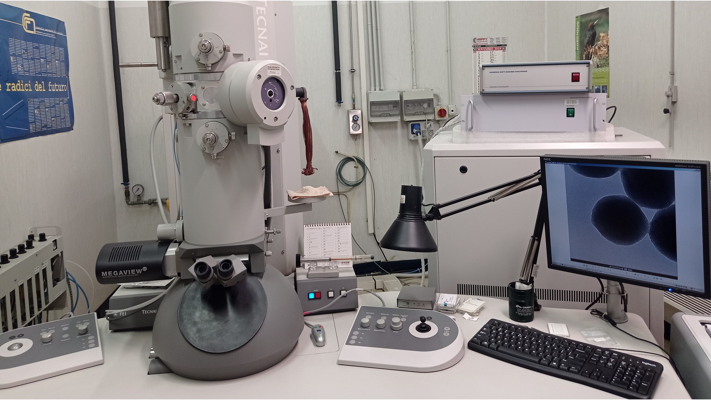
The FEI Tecnai G2 Spirit TWIN TEM at the CNR‑IPCB unit is a versatile transmission electron microscope designed for high‑resolution morphological and structural analysis of inorganic, organic, polymeric, and biological materials. It operates with a LaB₆ thermionic source and supports accelerating voltages up to 120 kV, enabling the observation of beam‑sensitive specimens while maintaining excellent image quality. The system offers a line‑to‑line resolution of 0.20 nm and a broad magnification range from 18× to 650,000×, allowing detailed investigation of nanoscale features, crystalline domains, interfaces, and internal morphologies. Imaging versatility is ensured by two digital cameras: a bottom‑mounted FEI Eagle 4k CCD for high‑sensitivity acquisition and a side‑mounted Olympus SIS MegaView G2 CCD for rapid imaging and diffraction pattern capture. The microscope supports bright‑field, selected‑area electron diffraction, and low‑dose imaging modes, making it suitable for both qualitative and quantitative structural studies. Sample preparation is enhanced by a LEICA EM UC6/FC6 cryo‑ultramicrotome, enabling the production of ultrathin cryo‑sections that preserve native structures in soft, hydrated, or biological materials. This capability is essential for polymers, tissues, vesicles, and complex soft‑matter systems.
Research areas and applications
The TEM FEI is very useful for the morphological and structural characterization of inorganic, organic and biological materials in bright field mode. Crio-ultramicrotomy for the preparation of ultrathin sections for TEM observations is also available.
Science highlights