TLM platform
General Information
Technique
Key Instrumentation
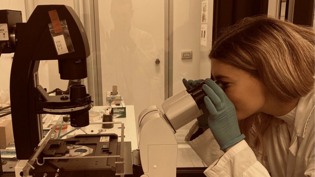
Time Lapse Microscopy for Organ on chip applicationsThe LEICA DMi8 inverted optical (brightfield) microscopy is utilized to capture time-lapse measurements of cells and tissues in lab-on-chip and organ-on-chip devices, employing an integrated incubator. Thanks to the optimal resolution, accuracy, and repeatability of the XYZ motorized stage, an unlimited number of regions inside the devices can be acquired to monitor, over time, more than one experimental condition. The top-stage incubator maintains the cell's physiological conditions throughout the experiment. Microfluidic inlets can ensure the integration of tubes for the automatic change of the medium inside the devices. The easy interface, realized in Python, allows the operator to choose the parameters for the experiment such as the desired regions of the chip, the exposure time, the total number of frames to acquire, and the interval between each frame.

Tecnical description
Utilizing inverted microscopy enables the acquisition of time-lapse measurements for dynamic studies involving cells and tissues within the innovative domains of lab-on-chip and organ-on-chip devices exploiting an integrated incubator. This advanced imaging technique facilitates meticulous observation and enhances the precision and depth of analysis, allowing for a comprehensive exploration of cellular and tissue behaviour in a controlled microenvironment. The inverted LEICA DMi8 microscope is characterized by an XYZ motorized stage with dimensions of (L x W x H) 375 mm x 330 mm x 27 mm, positioning range of 127 mm x 83 mm, resolution of 0,7 μm, accuracy < 20 μm, and repeatability < 3 μm. It includes magnifications between 5x and 40x and is equipped with a CMOS camera (18MP, MU1803 Amscope). The incubation system is a top-stage incubator (OKOLAB) characterized by a chamber that fits the microscope stage. It allows the maintenance of temperature at 37°C and CO 2 at 5% with two feedback modes: sample and chamber modes. The temperature accuracy is ± 0.1°C and ± 0.3°C respectively, in sample and chamber feedback mode. A total of 25 holes, located on two sides of the chamber, guarantee access to the devices for different-sized microfluidic tubes.
Research areas and applications
Biomedical research, drug testing, disease modelling
Science highlights
Experimental team

- Eugenio Martinelli
- University of Rome Tor Vergata
- Professor
