PET/CT, GE Discovery
General Information
Technique
Key Instrumentation
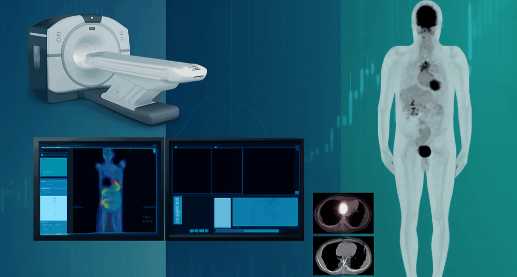
The instrument PET/CT is a GE Discovery MI 4R with SiPM located at the Unit TECNOMED in Monza. The equipment is designed to enhance diagnostic confidence and improve patient care in PET/CT imaging.
It is equipped with LightBurst Digital Detectors using SiPM technology paired with lutetium-based scintillators. It offers significantly improved sensitivity: ~13.3 cps/kBq (NEMA), nearly double that of equivalent PMT-based analog systems.
TOF resolution ~377 ps FWHM, enhancing lesion contrast and localization precision.
Diagnostic-quality CT integrated with performix 40 Plus X-ray tube, highlight detectors for enhanced resolution, ASiR-V iterative reconstruction, reducing CT dose up to 82% without compromising image quality. It supports clinicians in detecting disease earlier, assessing accurate treatment response, and making informed therapeutic decisions. By combining advanced digital technology with efficient workflows, it enables high-quality imaging with greater speed and reduced patient dose, contributing to a more comfortable and effective diagnostic experience. Its versatility makes it ideal for a wide range of clinical applications, including oncology, neurology, and cardiology.
Technical description
The 4-ring design provides 20 cm axial field of view (FOV) offers a strong balance between sensitivity and whole-body coverage, enabling efficient dynamic and total-body imaging protocols – reduced scan times. Spatial resolution at centre ~4–4.6 mm FWHM across radial, tangential and axial dimensions. High contrast recovery: 73–91% for spheres ranging from 10–37 mm; background variability of 1.8%. Delivers excellent detectability even for small lesions at low dose levels. The Peak NECR: ~176 kcps at 20 kBq/mL, outperforming earlier analog DMI platforms, enables faster scanning – lower injected activity, benefiting paediatric and high- throughput settings. Integrates TOF with Q.Clear (Bayesian Penalized Likelihood Reconstruction/BSREM) for higher image sharpness and SUV consistency provides improved lesion detectability, greater SUV accuracy, and up to 2x signal-to-noise improvement compared to OSEM.
Research areas and applications
In vivo imaging of PET Radiopharmaceuticals. The main clinical application are advanced oncology workflows, theranostic protocols, low-dose dynamic imaging, pediatric and whole-body PET and clinical research requiring high quantitative accuracy.
Science highlights
Chicheportiche, A., Marciano, R. & Orevi, M. Comparison of NEMA characterizations for Discovery MI and Discovery MI-DR TOF PET/CT systems at different sites and with other commercial PET/CT systems. EJNMMI Phys 7, 4 (2020). https://doi.org/10.1186/s40658-020-0271-x
Yoshii T, Miwa K, Yamaguchi M, Shimada K, Wagatsuma K, Yamao T, Kamitaka Y, Hiratsuka S, Kobayashi R, Ichikawa H, Miyaji N, Miyazaki T, Ishii K. EJNMMI Phys. 2020 Sep 11;7(1):56. https://doi.org/10.1186/s40658-020-00325-8. PMID: 32915344; PMCID: PMC7486353;
S, Basset-Sagarminaga J, van de Weijer T, Schrauwen-Hinderling VB, Backes WH, Wierts R. Improving image reconstruction to quantify dynamic whole-body PET/CT: Q.Clear versus OSEM. EJNMMI Phys. 2025 Mar 27;12(1):27. https://doi.org/10.1186/s40658-025-00736-5. PMID: 40140159; PMCID: PMC11947397
Mehranian, A., Wollenweber, S.D., Walker, M.D. et al. Image enhancement of whole-body oncology [18F]-FDG PET scans using deep neural networks to reduce noise. Eur J Nucl Med Mol Imaging 49, 539–549 (2022). https://doi.org/10.1007/s00259-021-05478-x
Wumener, X., Zhang, Y., Zang, Z. et al. The value of dynamic FDG PET/CT in the differential diagnosis of lung cancer and predicting EGFR mutations. BMC Pulm Med 24, 227 (2024). https://doi.org/10.1186/s12890-024-02997-9
Experimental team

- Università di Milano-Bicocca
Paola Anna Erba

- Università di Milano-Bicocca

- Università di Milano-Bicocca