Confocal Microscope 1
General Information
Technique
Key Instrumentation
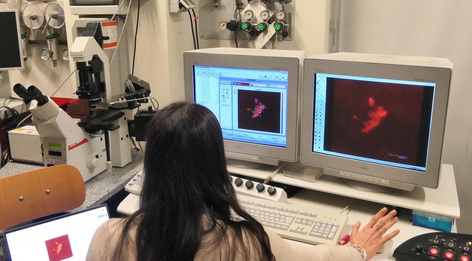
Laser Scanning Confocal MicroscopeA Laser Scanning Confocal Microscope Leica TCS SP2 is installed at the CSGI unit of the University of Florence, providing high‑resolution optical sectioning and 3D imaging for advanced studies in materials science, chemistry, biology and soft‑matter systems. The system is equipped with three fluorescence PMT detectors and a tunable spectral detection module, enabling precise separation of emission bands and simultaneous acquisition of multiple fluorophores. Its laser lines support excitation across the visible range, allowing efficient imaging of common dyes, fluorescent proteins and functionalized nanoparticles. The confocal scanning head offers high‑speed frame acquisition with diffraction‑limited lateral resolution, while the motorized Z‑axis enables accurate reconstruction of volumetric datasets and time‑lapse imaging of dynamic processes. The microscope includes a fully motorized stage for multi‑position experiments, tile scans and automated acquisition routines, ensuring reproducibility and stability during long imaging sessions. Advanced imaging modes—such as spectral unmixing, lambda scanning, FRAP and 3D reconstruction—expand the analytical capabilities of the platform, making it suitable for quantitative fluorescence studies, co‑localization analysis and structural characterization of complex samples. The system accommodates a wide range of objectives, from low‑magnification survey lenses to high‑NA oil‑immersion optics, supporting both large‑area mapping and nanoscale feature visualization. This configuration provides researchers and industrial partners with a versatile, reliable tool for high‑quality imaging of heterogeneous materials.
Technical description
Laser Scanning Confocal Microscope provides high‑resolution optical sectioning and quantitative fluorescence analysis for materials science, soft‑matter research and biological imaging. The system supports three‑dimensional imaging, spatially resolved emission mapping and advanced photophysical measurements, including FRET and FRAP, enabling users to investigate molecular interactions, diffusion processes and dynamic structural changes within complex samples. A transmission PMT extends detection capabilities to transmitted‑light modalities, improving contrast for non‑fluorescent or weakly labeled specimens. Eight excitation laser lines cover a broad spectral range, allowing efficient excitation of common fluorophores, fluorescent proteins and functionalized nanoparticles. The motorized microscope table enables fully automated 3D motion for large‑area imaging, multi‑position acquisition and long‑term experiments requiring precise spatial reproducibility. The optical configuration ensures diffraction‑limited lateral resolution and accurate Z‑stack reconstruction, supporting volumetric imaging of micro‑ and nano‑structured materials. Although it represents an entry‑level version compared to the Leica TCS SP8 platform, the instrument provides robust performance for routine confocal imaging, spectral separation, co‑localization studies and time‑lapse fluorescence analysis.
Research areas and applications
The instrument allows 3D chemical mapping of complex systems and interfaces; Electronics & Semiconductor, Automotive & Transportation; Metals & Machine Engineering; Medical Device QA/QC; Technical Cleanliness, Metallography, Material Analysis, Sample Preparation for Materials Science; live Cell Imaging, 3D Cell Culture.
Science highlights
- Biogenic Supported Lipid Bilayers from Nanosized Extracellular Vesicles, Montis et al. https://doi.org/10.1002/adbi.201700200
- Methylene blue-containing liposomes as new photodynamic anti-bacterial agents, Boccalini et al. https://doi.org/10.1039/C6TB03367A
- Magnetically Triggered Release From Giant Unilamellar Vesicles: Visualization By Means Of Confocal Microscopy, Nappini et al. https://doi.org/10.1021/jz2000936
Experimental team

- Marco Laurati
- CSGI-University of Florence
- Professor