TEM High Resolution
General Information
The Thermo Scientific Talos F200X STEM is a scanning transmission electron microscope that combines outstanding high-resolution STEM and TEM imaging with industry-leading energy dispersive X-ray spectroscopy (EDS) signal detection. 2D/3D chemical characterization with compositional mapping is performed by 4 in-column SDD Super-X detectors with unique cleanliness. The Talos F200X scanning transmission electron microscope allows for the fastest and most precise EDS analysis in all dimensions, along with high-resolution TEM and STEM (HRTEM and HRSTEM) imaging with fast navigation for dynamic microscopy.
The Talos F200X -(S)TEM delivers fast, precise, quantitative characterization of nanomaterials in multiple dimensions. With innovative features designed to increase throughput, precision, and ease of use, the Talos F200X- (S)TEM is ideal for advanced research and analysis in academic, government, semiconductor and industrial environments.
The need for large area correlative imaging at high resolution has recently increased as it allows researchers to preserve the context of their observations while also providing statistically robust data.
Thermo Scientific Map Software(enable by Thermo Scientific Velox Software) automatically acquires an array of images across a sample and stitches them together to create one large final image. Image acquisition can even be performed unattended.
This technique uses a series of 2D images, obtained at different tilt angles, to reconstruct a 3D model of the sample through computational algorithms.3D chemical mapping of complex system and interface.
Technical description
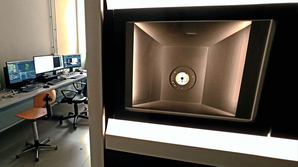
The Thermo Scientific Talos F200X at the CNR‑ICCOM unit is a 200 kV field‑emission S/TEM designed for high‑resolution imaging and advanced analytical characterization of inorganic, organic, and hybrid materials. Its FEG source provides a bright, stable beam suitable for both TEM and STEM modes, enabling HRTEM and HRSTEM imaging with sub‑nanometer detail. The system integrates four large‑area Super‑X EDS detectors arranged symmetrically around the sample, ensuring outstanding X‑ray collection efficiency and enabling fast, quantitative elemental mapping in two and three dimensions. This configuration supports high‑precision chemical analysis even at low beam currents, making it ideal for beam‑sensitive materials. The microscope offers rapid navigation and dynamic imaging capabilities, allowing real‑time observation of structural evolution, nanoparticle transformations, and other time‑dependent processes. Automated alignment routines and advanced drift‑correction tools ensure stable long‑duration acquisitions, essential for high‑resolution studies and tomography. The instrument supports a wide range of analytical techniques, including electron diffraction, STEM‑HAADF imaging for Z‑contrast analysis, and spectrum‑imaging workflows for correlative structural–chemical interpretation.
Research areas and applications
-Transmission electron microscopy (TEM) is a high-resolution imaging technique in which a beam of electrons passes through a thin sample to produce an image. The electron beam is impacted by the sample’s thickness/density, composition, and, in some cases, crystallinity. The electrons that are transmitted through the specimen subsequently provide contrast in the resulting image, revealing high-resolution compositional information.
-TEM is capable of obtaining data at virtually unparalleled resolution due to the small wavelength of the transmitted electrons. Unlike scanning electron microscopy (SEM), which gathers the net intensity of secondary electrons in each point of the scan, TEM resolution is only limited by the wavelength of the individual electron and the quality of the electron optics. This means that transmission electron microscopes can routinely collect sub-nanometer-resolution (or even atomic-resolution) images.
-HRTEM is used both in biology (for example, in the study of cellular ultrastructures) and in materials science for the study and characterization of microphases and interfaces.
– Electron tomography, powered by Thermo Scientific, delivers detailed 3D structural information at the nanometer scale. This technique uses a series of 2D images, obtained at different tilt angles, to reconstruct a 3D model of the sample through computational algorithms.
Science highlights
Pietro Mariani; Xiao Sun; Enrico Berretti; Alessandro Lavacchi; et al. Inorganic Chemistry Frontiers-(2024). <a href=”https://doi.org/10.1039/D4QI02391A”>https://doi.org/10.1039/D4QI02391A </a>
Francesca Liguori; Werner Oberhauser; Enrico Berretti; Lorenzo Poggini; Pierluigi Barbaro; Carmen Moreno‐Marrodán. Advanced Energy and Sustainability Research (2025). <a href=”https://doi.org/10.1002/aesr.202400349″>https://doi.org/10.1002/aesr.202400349</a>
Samuele Fanetti; Sebastiano Romi; Enrico Berretti; Michael Hanfland; Emin Mijit; Frederico Alabarse; Philip Dalladay-Simpson; Federico Gorelli; Roberto Bini; Mario Santoro -The Journal of Chemical Physics (2023). <a href=”https://doi.org/10.1063/5.0167748″>https://doi.org/10.1063/5.0167748</a>
Experimental team

- Alessandro Lavacchi
- CNR-ICCOM
- Researcher

- Maria Cristina Salvatici
- CNR-ICCOM
- Researcher

- Beatrice Muzzi
- CNR-ICCOM
- Staff Researcher