AFM BIO
The BIO Atomic Force Microscope (AFM) is a high-resolution scanning probe microscope dedicated to the study of biological and soft-matter samples at the nanoscale. The BIO AFM is designed specifically for biological applications, allowing imaging and mechanical characterization of living cells, biomolecules, and other delicate samples under near-physiological conditions.
It uses a sharp nanometric tip mounted on a flexible cantilever to scan sample surfaces, producing three-dimensional topographical maps with nanometer resolution. Unlike conventional AFMs optimized for dry samples, BIO AFM can work in fluid, enabling observation of living cells, membranes, proteins, extracellular vesicles, and other hydrated biological structures. Thanks to low interaction forces, it allows investigation of fragile specimens such as soft tissues, biofilms, and macromolecular complexes.
The instrument can measure physical properties including elastic modulus (stiffness), adhesion forces, viscoelastic behavior and surface roughness. In particular, it is commonly used for imaging living cells and cellular membranes, studying protein aggregates and DNA structures, characterizing biomaterials and nanomedicine systems, investigating extracellular vesicles and mineralization processes, measuring interactions between biomolecules and surface analysis of biocompatible materials. Atomic force microscopy is particularly valuable in biomedical research because it can reveal structural and mechanical information that complements optical and electron microscopy.
Within the NAST infrastructure, the BIO AFM contributes to interdisciplinary projects spanning from nanomedicine to biomaterials.
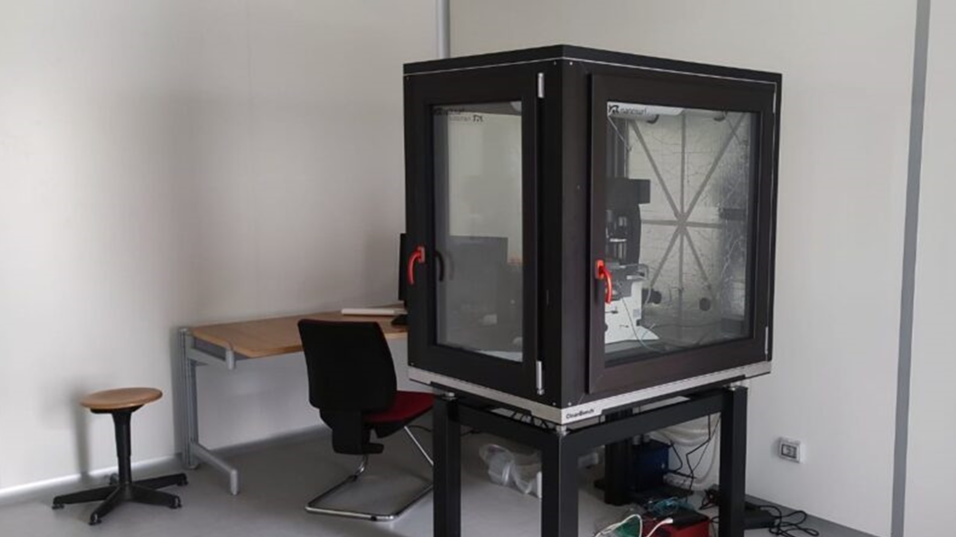
Technical description
This BIO Atomic Force Microscope (AFM) allows to characterise of a maximum lateral dimension of 100 µm x 100 µm, a maximum height of 10 µm, and it is equipped with a microfabricated cantilever – for conductometry measurements – that scans the sample surface. Interatomic forces(the force sensitivity of this instrument is pico-Newton range) cause cantilever deflection, which is detected by an optical beam-deflection system (laser + photodiode). A piezoelectric scanner provides sub-nanometer positioning in X-Y-Z axes: the scan speed is adjustable (seconds to minutes per image). This mechanism enables topographic imaging with nanometer lateral resolution and sub-nanometer vertical resolution. The optical microscope features magnification factors of 5x, 10x, 20x, 40x and maximum movements in the XY stage of 10 mm x 10 mm. This BIO Atomic Force Microscope (AFM) operational modes include:
• non-contact mode, useful for delicate structures. Tip oscillates above the surface detecting attractive forces;
• solid/liquid contact mode, suitable for rigid samples in order to measure friction and lateral forces. Tip maintains constant force on the surface;
• solid/liquid tapping mode, preferred for soft and living biological materials. Cantilever oscillates near resonance. Thank to this mode, AFM minimizes sample damage;
• solid/liquid with force spectroscopy;
• solid/liquid nanolithography.Research areas and applications
Topographical analysis pf solid substrates and biological samples in liquid, nanolithography of rigid substrates, nanostimulation of cells.
Science highlights
Experimental team

- Università di Roma Tor Vergata
- Professor