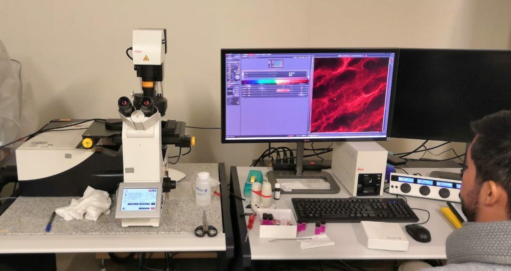
Confocal Microscope 2
General Information
Technique
Laser Scanning Confocal Microscope Leica TCS SP8 equipped with DMI8 microscope, FCS Picoquant module and with PMT detector for transmission imaging and 5 internal detectors.

Tecnical description
The Laser Scanning Confocal Microscope is a Leica TCS SP8, equipped with DMI8 microscope, FCS Picoquant module and with PMT detector for transmission imaging and 5 internal detectors. The latter are two PMT, one hybrid detector and two cooled hybrid detectors, suitable for Single Molecule Detection, 3D imaging, spatially resolved imaging, and spatially resolved FRET, FRAP, FCS and FCCS. The lasers available allow 8 excitation lines, and the microscope table allows motorized motion in 3D for imaging of larger samples. Compared to the Leica TCS SP2, this instrument is recommended when FCS and FCCS analysis are required.
Research areas and applications
The instrument allows users to perform 3D chemical mapping of complex systems and interfaces; Electronics & Semiconductor, Automotive & Transportation; Metals & Machine Engineering; Medical Device QA/QC; Technical Cleanliness, Metallography, Material Analysis, Sample Preparation for Materials Science; live Cell Imaging, 3D Cell Culture. In particular, the FCS and FCCS options are relevant samples of biological interest, where dynamics and diffusion processes are investigated.
Science highlights
- Dewetting acrylic polymer films with water/propylene carbonate/surfactant mixtures – implications for cultural heritage conservation, M. Baglioni et al. https://doi.org/10.1039/D1CP03201A
- Interaction of nanoparticles with lipid films: the role of symmetry and shape anisotropy, L. Caselli et al. https://doi.org/10.1039/C7CP02608K
- Organized Hybrid Molecular Films from Natural Phospholipids and Synthetic Block Copolymers: A Physicochemical Investigation, A. Balestri et al. https://doi.org/10.1021/acs.langmuir.0c01544
- Microgel dynamics within the 3D porous structure of transparent PEG hydrogels, Bassu et al. https://doi.org/10.1016/j.colsurfb.2022.112938
Experimental team

- Marco Laurati
- CSGI-University of Florence
- Professor
