Raman Confocal Microscope
General Information
Unit
CSGITechnique
Key Instrumentation
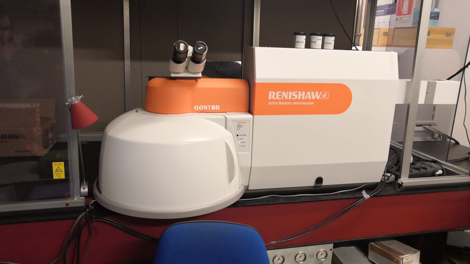
The Raman Confocal Microscope, available at the CSGI – University of Florence unit, is a Renishaw inVia™ Qontor™ system designed for high‑performance vibrational spectroscopy and chemical imaging across a wide range of research fields.
The instrument is equipped with two excitation wavelengths, 532 nm and 785 nm, enabling optimized analysis of both highly scattering materials and samples prone to fluorescence. Its fully motorized XYZ stage ensures precise and reproducible positioning, supporting automated mapping, multi‑area acquisitions, and long‑duration experiments with high spatial accuracy. The system integrates advanced confocal optics that provide sub‑micrometric lateral resolution and efficient rejection of out‑of‑focus light, allowing detailed characterization of heterogeneous materials, thin films, biological specimens, and micro‑structured surfaces. A flexible sampling arm extends the microscope’s capabilities to large, irregular, or non‑transportable samples, enabling in‑situ Raman measurements directly on objects of complex geometry. The platform supports a wide range of Raman imaging techniques, including point‑by‑point mapping, line scanning, and StreamLine™ fast imaging, facilitating rapid acquisition of chemically resolved images with minimal sample exposure. Automated calibration routines, high‑stability optics, and integrated software for spectral processing and multivariate analysis ensure reliable data quality and efficient workflow management.
Overall, the inVia™ Qontor™ Raman Confocal Microscope provides a versatile and powerful solution for researchers and industrial users requiring precise molecular‑level characterization and high‑resolution chemical imaging.
Technical description
The Raman Confocal Microscope is a Renishaw inVia™ Qontor™ system designed for high‑precision vibrational spectroscopy and chemical imaging. It operates with two excitation wavelengths, 532 nm and 785 nm, enabling optimized analysis of a wide variety of materials, including those affected by fluorescence or requiring deeper penetration.
The instrument features a fully motorized XYZ stage that ensures accurate sample positioning and supports automated Raman mapping with high spatial reproducibility. A flexible sampling arm extends the system’s capabilities to large or irregular objects, allowing in‑situ measurements on samples that cannot be placed directly under the microscope. The platform integrates advanced confocal optics for sub‑micrometric resolution and efficient rejection of out‑of‑focus light, making it suitable for heterogeneous materials, thin films, biological specimens, and microstructured surfaces.
A key component of the system is the LiveTrack™ technology, which enables real‑time focus tracking during spectral acquisition and Raman imaging. This capability is essential for analyzing samples with coarse, uneven, or sloping surfaces, ensuring consistent focus and reliable data quality throughout long or complex mapping sequences. The microscope supports multiple Raman imaging modes, including point‑mapping, line scanning, and fast imaging, and is complemented by robust software for spectral processing and multivariate analysis.Research areas and applications
3D chemical mapping of complex systems and interfaces
Science highlights
Experimental team

- CSGI
- Professor
Emiliano Fratini

- CSGI
- Professor