F. Tavola, G. P. De Gaudenzi, G. Bidinotto, F. Casamichiela, A. Pola, S. Tedeschi, B. Bozzini. ChemSusChem. 18(10), e202402218 (2025). doi:10.1002/cssc.202402218.
RETINA
General Information
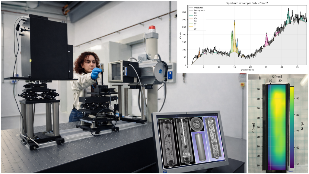
RETINA is a laboratory-based X-ray platform designed for non-destructive elemental analysis and imaging of medium-sized samples (up to 15 cm). It supports qualitative X-ray fluorescence (XRF), routinely detecting elements from Ca to Pb. With an accurately characterized tube spectrum and a thin, certified XRF calibration standard, RETINA can also perform quantitative XRF on planar specimens, especially when thickness is limited and the measurement geometry is well defined. A high-precision XYZ positioning system enables repeatable raster scans over centimeter-scale areas, while an adjustable collimator defines the excitation spot and allows 2D elemental mapping with millimeter-scale lateral resolution (step size and collimation dependent). Radiographic imaging is available in the same setup, providing fast sample inspection and alignment before or during XRF measurements. RETINA is equipped with an X-ray imaging detector offering a field of view up to 25 cm, enabling radiography and cone-beam CT of medium-sized objects. Depending on sample size, geometry and acquisition settings, the minimum achievable spatial resolution in reconstructed data can reach ~80 μm. The combination of XRF mapping and imaging makes the instrument well suited to study composition–structure relationships, from planar energy materials (e.g., PEMFC membranes and battery electrodes) to volumetric inspection and tomography of compact devices (e.g., sealed battery cells, electronic components) and other heterogeneous objects requiring a flexible, laboratory-based setup. User-selectable collimation and scan strategies provide a practical trade-off between sensitivity, resolution and throughput.
Technical description
RETINA is equipped with a high-power X-ray tube (IAE RTC1000HS) operated from 40 to 150 kV. The anode current is up to 5 mA in continuous mode or 200 mA in pulsed mode, providing an integral fluence at 1 m of ~10^8 and ~10^10 photons cm−2 s−1, respectively. Samples are handled by a high-precision THORLABS positioning system with three motorized translation axes (XYZ) and a vertical rotation axis, enabling both raster scans for XRF mapping and cone-beam CT acquisitions. The excitation spot is defined by adjustable/interchangeable collimation, typically supporting millimetre-scale mapping (step size and collimator dependent). XRF spectra are acquired with a Peltier-cooled CdZnTe detector (Amptek XR-100T-CZT), covering ~3–90 keV with ~0.4 keV FWHM at Fe Kα; spectra are processed in PyMCA using a fundamental-parameters approach, supported by a characterized tube spectrum and thin calibration standards. Imaging is performed with a large-area X-ray detector (up to 25 cm field of view) for 2D radiography and 3D tomography; depending on geometry and sample size, reconstructed spatial resolution can reach 80 μm. The system supports flexible source–sample–detector distances to balance magnification, flux and field of view. Tube and motion control are integrated to synchronize stage motion and detector live time, enabling automated multi-point acquisitions with repeatable geometry.
Research areas and applications
RETINA enables integrated XRF mapping and X-ray imaging/tomography to study composition–structure relationships across energy materials, geoscience and heritage/industrial specimens. A core line of research targets electrochemical systems and degradation mechanisms: PEM fuel-cell components (membranes, GDLs and catalyst layers), battery electrodes and full cells, and porous metallic/current-collector architectures. In these studies, 2D elemental maps are used to locate critical inhomogeneities (e.g., catalyst distribution, contaminant accumulation, corrosion products). Recent work also includes the development and validation of advanced CT workflows for dose-limited experiments, including hybrid acquisition strategies and instance-specific denoising for improved reconstruction quality.
RETINA is routinely applied to quantitative/qualitative XRF on planar samples (materials screening, process control, failure analysis) and to 3D inspection of medium-sized objects (sealed battery cells, engineered parts, heterogeneous composites). Beyond energy materials, the facility supports applied elemental investigations such as chromium quantification in leather (compliance and conservation studies) and the elemental/metallic composition analysis of fossilized bones, where combined mapping and imaging help distinguish mineral phases, diagenetic alteration patterns and metal enrichment.
Science highlights
Experimental team
Instrument Scientist

- Andrea Pola
- DENG-POLIMI
- Professor
Staff

- Francesco Casamichiela
- DENG-POLIMI
- IT

- Aixeen Manuel Fontanilla
- Politecnico di Milano
- PhD student