TESCAN UniTOM HR
General Information
The TESCAN UniTOM HR instrument enables multi-scale X-ray imaging and micro-CT with spatial resolution ranging from tens of microns down to the sub-micron level (down to 600 nm, depending on geometry and acquisition settings). It supports continuous 360° rotation and is designed for in situ and time-resolved experiments, enabling repeated scans and time-lapse tomography for dynamic studies.
The system accommodates large and heavy specimens (up to 300 mm in diameter, 400 mm in height, and 45 kg in weight) and features multiple detectors, balancing field of view, contrast and temporal resolution. This makes the platform suitable for multi-scale workflows, from overview scans of large objects to high-resolution region-of-interest imaging, while keeping a stable rotation and repeatable positioning for longitudinal studies.
In materials science, UniTOM HR is employed to study microstructures, porosity, cracks, and phase distribution in metals, ceramics, and composites. It plays a key role in additive manufacturing, supporting non-destructive quality control and defect detection in 3D-printed parts, including lattice structures and internal channels.
In the geosciences, it enables analysis of rock pore networks, mineral inclusions, and fossil structures, supporting studies of reservoir properties, connectivity and fluid flow. In battery research, it is used to investigate electrode degradation, dendrite formation, and microstructural changes during charging and discharging.
In polymer science, it aids in analysing foams, fibres, and fracture behaviour. Finally, in civil engineering it is used to assess concrete microstructure, crack formation, and moisture transport.
Technical description
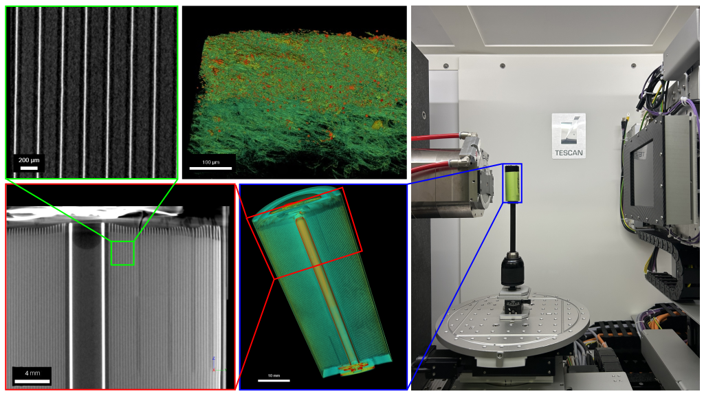
TESCAN UniTOM HR is a laboratory micro-CT system equipped with a 30–160 kV, 50 W open X-ray source and two flat-panel detectors: a low-energy panel (50 µm pixels) for high-contrast imaging at lower kV, and a second panel (75 µm pixels) optimized for higher energies and fast, low-dose acquisitions. The system provides continuous 360° rotation for true in situ and time-resolved tomography, allowing repeated scans during mechanical, thermal or electrochemical experiments. Multiple source focusing modes allow users to trade off spot size, flux and penetration; sub-micron imaging is achievable, with a best spatial resolution of ~600 nm under optimized conditions. The maximum imaging envelope is 400 mm (H) × 300 mm (Ø), supporting both large-object scans and high-resolution region-of-interest workflows via geometrical magnification and zoom reconstructions. Integrated software supports automated acquisition, reconstruction (FDK based, full and 180+ scans), and 4D analysis, including registration, temporal differencing and quantitative morphometrics. The control environment allows full-object scans as well as scripted multi-ROI “zoom” scans in a single session, ensuring consistent geometry and metadata for reproducible studies. Real-time radiography supports alignment; the stage accommodates custom in situ rigs and loads up to 45 kg. Data are logged for traceability.
Research areas and applications
X-ray micro-CT enables non-destructive 3D characterization across materials science, geosciences, life sciences and cultural heritage. This TESCAN UniTOM HR system is particularly suited to time-lapse and continuous dynamic scanning, allowing real-time observation of sample evolution and quantitative 4D analysis during in situ loading, heating or electrochemical operation. In materials science it measures microstructure, porosity, cracks and phase distribution in metals, ceramics and composites, and it is widely used for additive-manufacturing quality control and defect detection in 3D-printed parts (internal channels, lattices). In geoscience and palaeontology it resolves rock pore networks, mineral inclusions and fossil structures to support reservoir-property and fluid-flow studies. In battery research it tracks electrode degradation, dendrite formation and microstructural changes during cycling. In life sciences it images bone and soft-tissue morphology, small animals/plants and vascular networks without sectioning; in pharma it characterizes tablets, coatings and drug-delivery structures. For archaeology and conservation it reveals the internal build and damage of artefacts without sampling. Additional use cases include polymers (foams, fibres, fracture) and civil engineering (concrete microstructure, cracking, moisture transport) and non-destructive testing of objects.
Science highlights
F. Verducci, L. Cultrera, E. Colombo, A. M. Fontanilla, F. Casamichiela, D. Mazzucconi, A. Pola, A. Casalegno, A. Baricci. Journal of Power Sources. 660, 238538 (2025). doi:10.1016/j.jpowsour.2025.238538.
F. Tavola, G. P. De Gaudenzi, G. Bidinotto, F. Casamichiela, A. Pola, S. Tedeschi, B. Bozzini. ChemSusChem. 18(10), e202402218 (2025). doi:10.1002/cssc.202402218.
F. Gatti, F. Mirani, D. Mazzucconi, A. Pola, M. Passoni, IEEE Transactions on Instrumentation and Measurement, vol. 73, pp. 1-12, 2024, Art no. 3536912, https://doi.org/10.1109/TIM.2024.3472852
R. Ferretti, A. Casado Coscolla, F. Casamichiela, A. Pola, E. Vendrell Vidal, C. Sanchez Belenguer. IEEE Transactions on Computational Imaging. submitted (year n/a). DOI pending.
Experimental team

- Andrea Pola
- DENG-POLIMI
- Professor
- Davide Mazzucconi
- DENG-POLIMI
- Researcher

- Francesco Casamichiela
- DENG-POLIMI
- Technician