Confocal Microscope 3
General Information
Technique
Key Instrumentation
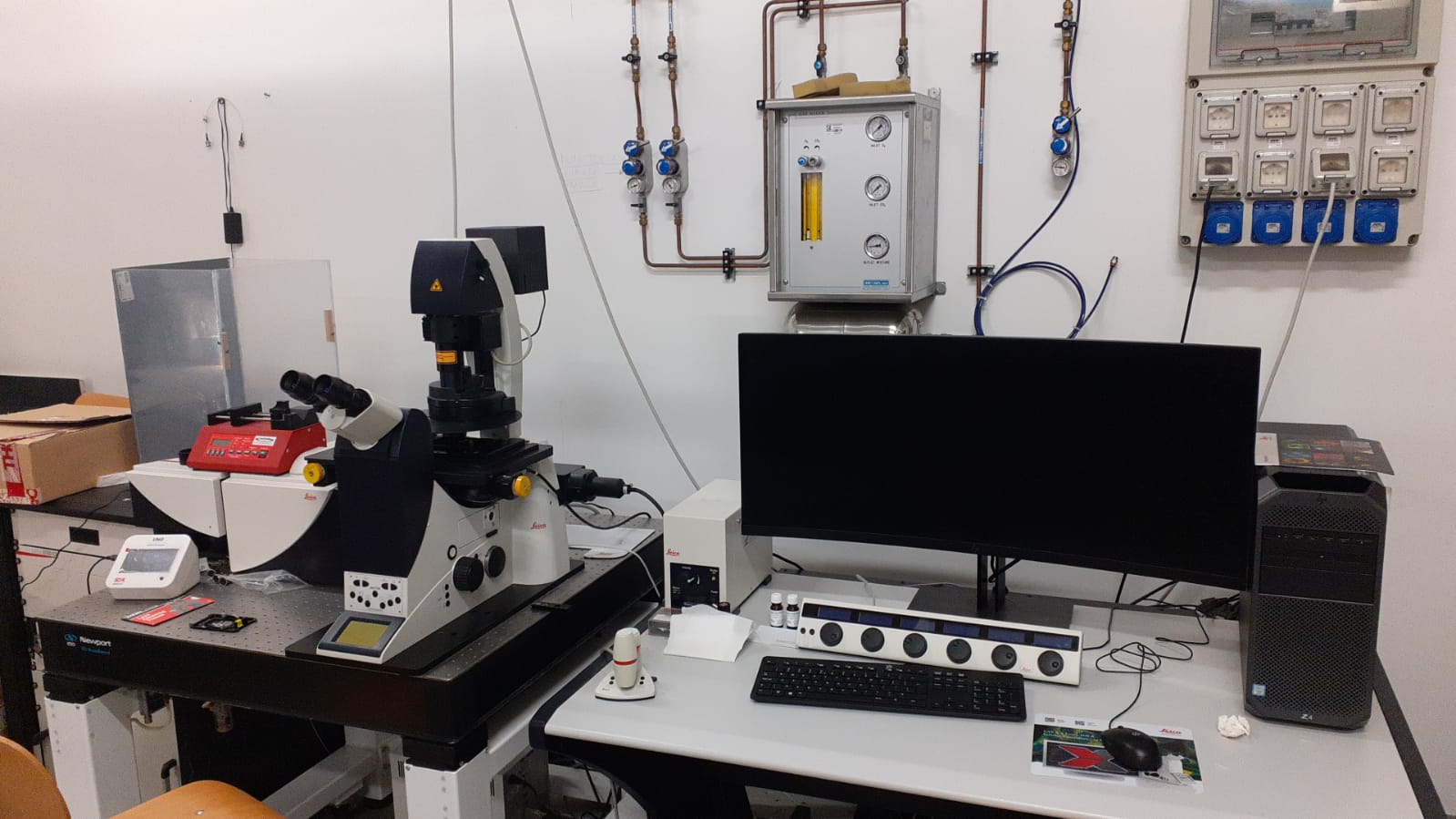
Leica TCS SP5 II FluorescenceA Leica TCS SP5 II Fluorescence Confocal Microscope is available at the University of Milano‑Bicocca, providing high‑sensitivity optical sectioning and multidimensional fluorescence imaging for advanced research in life sciences, soft matter, and functional materials. The system is built around a high‑speed resonant scanner and a conventional galvanometric scanner, enabling both rapid acquisition for live‑cell studies and high‑resolution imaging for detailed structural analysis. Its spectral detection architecture allows flexible selection of emission bands, supporting simultaneous imaging of multiple fluorophores with minimal crosstalk. The microscope is equipped with a suite of visible and near‑UV laser lines for efficient excitation of dyes, fluorescent proteins, and nanostructured probes. A tunable acousto‑optic beam splitter ensures precise control of laser power and wavelength routing, while hybrid and photomultiplier detectors provide high quantum efficiency and low noise for weak‑signal applications. The motorized XYZ stage enables automated tile scans, multi‑position experiments, and volumetric imaging with accurate spatial reproducibility. Advanced acquisition modes include lambda scanning, spectral unmixing, FRAP, FRET, and 3D reconstruction, supporting quantitative studies of molecular interactions, diffusion processes, and dynamic cellular responses. The system accommodates a broad range of objectives, from long‑working‑distance lenses for thick samples to high‑NA immersion optics for sub‑micrometric resolution. This configuration offers researchers and industrial partners a platform for high‑quality fluorescence imaging.
Technical description
For Leica TCS SP5 II Fluorescence Confocal Microscope, excitation is delivered through laser lines at 454, 488, 514 and 635 nm, enabling efficient stimulation of a wide range of fluorophores and fluorescent proteins. The detection system is equipped with Hybrid PMT detectors, offering high quantum efficiency, low noise and fast response, which makes the instrument suitable for weak‑signal imaging and rapid dynamic processes. Spectral resolution is available directly on the acquired images through a tunable detection module that allows flexible selection of emission bands and precise separation of overlapping spectra. The microscope also integrates a STED super‑resolution module, enabling sub‑diffraction imaging for structural studies requiring nanoscale detail. Its scanning architecture supports both high‑speed and high‑resolution acquisition, while the motorized XYZ stage ensures accurate positioning for 3D imaging, multi‑location experiments and large‑area tile scans. A broad set of objectives, including high‑NA immersion lenses, allows users to adapt the optical configuration to thick specimens, live‑cell imaging or nanoscale structural analysis. This combination of excitation flexibility, sensitive detection and super‑resolution capability makes the TCS SP5 II a robust platform for researchers and industrial users who require precise, quantitative fluorescence imaging.
Research areas and applications
Biomedical and cellular biology research.
Science highlights
Experimental team

- Maddalena Collini
- University of Milano Bicocca
- Professor

- Laura Sironi
- University of Milano Bicocca
- Laura D’Alfonso
- University of Milano Bicocca
- Professor