Fluorescence Microscopy
General Information
Technique
Key Instrumentation
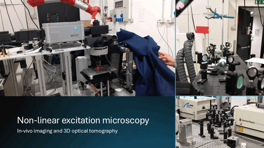
Nonlinear excitation fluorescenceThe facility is devoted to the investigation of the structure, function, and dynamics of biological systems through advanced non‑linear excitation microscopy. Its mission is to provide researchers with state‑of‑the‑art tools capable of probing living tissues, cellular architectures, and biomolecular processes with high spatial and temporal resolution. At the core of the laboratory is a direct raster‑scanning microscope designed for neurophysiology, which is coupled to two independent femtosecond laser sources. This dual‑laser configuration enables flexible excitation schemes and supports optical tomography of thick and highly scattering specimens, allowing users to explore biological samples well beyond the limits of conventional linear microscopy.
The facility offers both two‑photon fluorescence microscopy and polarization‑resolved second harmonic generation (SHG), two complementary techniques that together cover a broad range of biological questions. Two‑photon excitation can be applied to fluorescent proteins such as GFP and its engineered mutants, as well as to intrinsic metabolic cofactors like NADH and FAD, enabling label‑free or minimally invasive imaging of cellular metabolism. Time‑resolved digital detection, combined with the use of a SPAD array, allows full FLIM (fluorescence lifetime imaging microscopy) analysis, providing quantitative information on metabolic states, redox balance, and calcium dynamics within living cells and tissues. In parallel, SHG and polarization‑dependent SHG microscopy offer a powerful, non‑invasive approach to studying highly ordered biological structures. Custom sample holders can be deigned and fabricated via 3D laser printing.
Technical description
The facility investigates the structure and dynamics of biological systems using advanced non‑linear excitation microscopy. The instrument is built on an Olympus BX51 main frame coupled to an ISS M620 scanning head (Urbana‑Champaign, IL). Excitation is provided by a DeepSee femtosecond laser (690–1040 nm tuning range, 80 MHz rep rate, ~70–100 fs pulse width at the cavity exit) with full GVD compensation, enabling optical tomography of thick specimens. The system supports two‑photon fluorescence microscopy and polarization‑resolved SHG. Three detection channels with multiple bandpass filters allow selective acquisition of GFP variants and SHG signals in both scanned and descanned modes. In descanned configuration, a 7×7 SPAD array (Genoa Instruments) enables ISM with state‑of‑the‑art reconstruction algorithms. Application of custom algorithms allows to single out the out-of-focus contribution from the in focus contribution. This is particularly suited to study samples endowed with low-medium scattering coefficients. Time‑resolved detection supports FLIM for metabolic imaging (NADH, FAD) and calcium dynamics, suited for neuro-imaging. SHG and polarization‑dependent SHG provide detailed analysis of collagen fibril organization in tissues. For applications in highly turbid media, phase reversal correction based on digital holography can be implemented on a parallel system.
Research areas and applications
Biomedical Research, Imaging of tissue and cells, Nanoscopy for biotechnology and Medicine (Collini, Chirico), Stochastic Simulations for Biophysics (Chirico), GFP mutants for biotechnology (Collini), Nanoparticles for Biomedical Applications (Chirico, D’Alfonso), In-vivo non-linear microscopy for biotechnology and Medicine (Sironi)
Three main areas: Biomedical Research, Nanoscopy and Physical Optics, Nanofabrication.
A. Biomedical Research (main area).
A1. Imaging of tissue and cells with confocal and non.-linear excitation microscopy, mainly based on two photons excitation fluorescence (TPEF) and second harmonic generation or scattering (SHG) imaging. Through three acquisition channels tuned on GFP proteins and its mutants we follow the expression of tagged proteins. Through quantum dots and gold anistropic nanoparticles we follow endosomes kinetics in cell cytoplasm.
A2. We develop polarization dependent SHG imaging with phasor analysis and Machine Learning algorithms for clustering of features on the tissue image. Via fluorescence lifetime imaging with phasor based analysis we single out multispecies dynamics.
A3. We develop methods for data analysis encompassing image restoration and signal/noise ratio improvement. Virtual staining algorithms are also active area of research.
A4. We develop and test Image scanning microscopy for improved spatial resolution, with possibility to follow antibunching
A5. In vivo imaging on the same microscope with a thermostatted chamber and facility for small animal anesthesia.
B. Nanoscopy and Physical Optics.
B1. algorithms for the tissue scattering correction based on TPEF ;
Science highlights
Experimental team

- Giuseppe Chirico
- University of Milano Bicocca
- Professor

- Maddalena Collini
- University of Milano Bicocca
- Professor
- Laura D’Alfonso
- University of Milano Bicocca
- Professor

- Laura Sironi
- University of Milano Bicocca