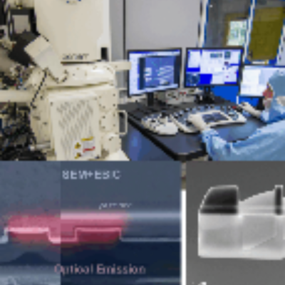
EcoBiM LAB (that is “Microbial Ecology and Biotechnology LAB”) is dedicated to the preparation of environmental samples and to the extraction, quantification, and quality control of microbial DNA from a wide range of environmental matrices, including seawater, sea ice and snow, soil, plant roots and marine sediments. The facility provides standardized workflows and instruments to ensure high-quality nucleic acids for downstream molecular ecology, microbiome and microbial genomics
ESCALAB QXi is a multi-technique photoelectron spectrometer supporting XPS, UPS, REELS and ISS, with a dual mode ion gun for monoatomic or cluster beams. It ensures the production of high-quality DNA suitable for a broad spectrum of downstream molecular applications. A charge compensation system enables XPS on insulating samples. This facility features two detector setups: six channel arrays for spectroscopy, a parallel imaging system with channel plates and a position sensitive detector
The FIB-SEM (Focused Ion Beam – Scanning Electron Microscope) system offers high-resolution imaging and characterization of the sample surface. The focused Gallium ion beam is a versatile tool for precise milling of the bulk sample, allowing exposure of sub-surface details or fabrication of site-specific Transmission Electron Microscopy specimens. The FIB-SEM is also used for nanofabrication as the pattern generator and injection system for precursors allows the fabrication of complex structures
The DualBeam in systems aligns the focal points of the electron and gallium ion beams, optimizing a wide range of applications. This configuration enables simultaneous Scanning Electron Microscope imaging, during Focused Ion Beam milling, greatly improving precision, performance, and throughput in operations that require highly accurate material modification. The flexibility of this instrument is unique to TESCAN and will provide best-in-class accuracy and throughput for EBSD and 3D-EDS
The Biophotonics group develops new optical nonlinear microscopy methods for in vivo biophysics and medical physics. In this field the group is developing two photon fluorescence imaging and second harmonic generation microscopy. One of the main application aims to the study of is studying lymphocyte motion in lymph nodes, in order to model immune responses in mice. This work is being carried out thanks to a collaboration with the Biotechnology group in Milano – Bicocca University
The Nicolet Nexus 870 is an Fourier Transform infrared spectroscopy and microscopy system with an external optical table, MCT and TRS external detectors, a GeATR unit, a Hind photoelastic modulator, and one infrared continuum microscope. It enables simultaneous imaging and Fourier Transform infrared analysis of micrometric particles or domains, supporting chemical mapping of flat samples and powders and it is a fast technique for material quantification of constituents in a sample
The Nicolet iS10 is a Fourier Transform infrared spectrometer designed to identify unknown contaminants, analyze failures and characterize mixtures, using the high performance LightDrive optical engine components (a source, a laser, an interferometer and a detector). This instrument offers high spectral resolution (better than 0.25 cm-1) and signal to noise ratios (50 000:1) for detecting small defects. Performance is verified with an integrated validation wheel, using certified standards
Gas chromatograph (GC) is coupled to mass spectroscope (a single quadrupole) for identifying and quantifying chemicals in mixtures. The instrument is complemented by an autosampler, that can accommodate up to 40 samples and enabling automatic analysis and equipped with temperature control. The Gas Chromatography Ion Mobility Spectrometer QP2020NX model, from Shimadzu in Kyoto, Japan, is suitable for general untargeted analysis, as well as for the detection of specific chemical families
Thermo Fisher Scientific Delta V Advantage isotope ratio mass spectrometer – equipped with GasBench II, Flash 2000 Organic Elemental Analyzer and Trace GC Ultra – enables the determination of relative quantities of stable isotope ratios of C, H, O, N and S in solid and liquid non-volatile samples and in single compounds separated through gas chromatography. All the facility components (IRMS, GC and EA) are connected through the Thermo Scientific ConFlo IV Universal Interface
Raman spectroscopy is a non-destructive chemical analysis technique, used in materials science to study a substance’s unique molecular vibrations and crystal lattice structure. Raman imaging is a powerful technique that combines Raman spectroscopy with spatial resolution to analyze material properties. The LabRAM Odyssey is equipped with a spectrometer, to achieve high spectral resolution, and with an optical microscope with a high magnification objective